Purkinje neurons are unique cells that are critical elements within the complex mechanism of our brain. Located within the cerebellum, they possess distinct morphological features that make them vital to our body’s ability to move harmoniously. Here we look at the anatomy of Purkinje neurons, their essential role in motor coordination, and how they influence our ability to learn new motor skills and adapt to changes.
Contents
Introduction to Purkinje Neurons
In the vast realm of neuroscience, one particular type of neuron stands out due to its pivotal role in motor coordination: the Purkinje neuron. These neurons are central to our understanding of the complex interplay of brain structures involved in movement.
Definition of Purkinje Neurons
Named after Jan Evangelista Purkyně, a pioneering Czech anatomist and physiologist, Purkinje neurons are large cells located in the cerebellar cortex of the brain. They are known for their distinct branching dendritic tree, a structure that allows them to receive and process a significant amount of information. Notably, Purkinje neurons are the only neurons in the cerebellar cortex that send information out of the cerebellum to other parts of the brain.
Brief History of Purkinje Neurons’ Discovery
The discovery of these neurons is attributed to Purkyně, who first described them in 1837. His work marked an important advance in our understanding of the brain’s complexity. Purkyně’s intricate descriptions of the neuron’s structure and location within the cerebellum laid the foundation for further study.
Over time, these neurons have become the focus of extensive research due to their unique characteristics and critical role in motor coordination. They continue to be a rich source of insights for neuroscientists aiming to unravel the complex mechanics of the human nervous system.

Anatomy of Purkinje Neurons
Understanding the role of Purkinje neurons in motor coordination requires a deep dive into their anatomy. This includes their specific location within the brain, their unique morphological features, and the synaptic connections they form with other cells. The intricacies of their structure are as fascinating as they are fundamental to their function.
Location within the Brain
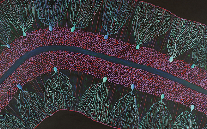
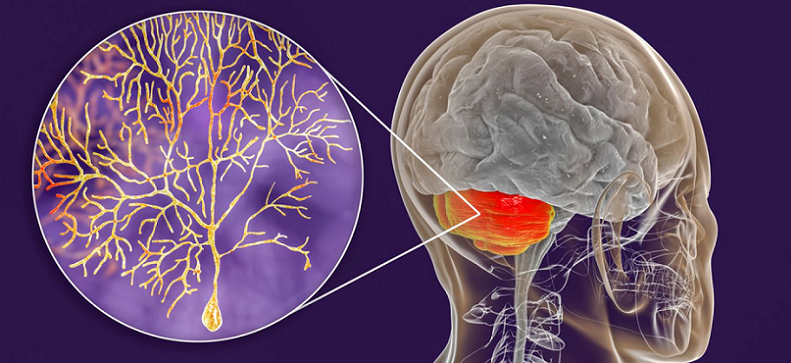
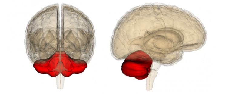
Purkinje neurons are situated in the cerebellar cortex, a layer of tissue at the surface of the cerebellum. The cerebellum, often referred to as the “little brain” due to its distinctive shape, is located at the back of the skull, underneath the cerebral hemispheres. Within the cerebellar cortex, Purkinje neurons align themselves in a single row, forming what is known as the Purkinje layer. Each neuron extends a large dendritic tree into the molecular layer above, while its axon passes through the granule cell layer beneath, eventually leaving the cerebellum [1].
Unique Morphological Features
Purkinje neurons are distinguished by their extensive and highly branched dendritic arbor, which appears as a complex, tree-like structure when viewed under a microscope. This unique morphology allows a single Purkinje cell to receive and integrate signals from a large number of inputs, up to 200,000 in some cases.
Additionally, these neurons have a large, flask-shaped soma, or cell body, from which the dendritic tree emerges. Their axons are also notable for being the only efferent, or output, axons from the cerebellar cortex, highlighting their critical role in transmitting the cerebellum’s outputs to other brain areas.
Synaptic Connections
Purkinje neurons receive two major types of synaptic inputs: from parallel fibers and from climbing fibers. Parallel fibers are the axons of granule cells, which run horizontally across the cerebellar cortex and make contact with numerous Purkinje cells along their path. Climbing fibers, on the other hand, originate from neurons in the inferior olive, a brainstem nucleus. Each climbing fiber forms an intimate connection with a single Purkinje cell, wrapping around its dendritic tree and providing a powerful excitatory input [2].
These connections create a dense network of communication, allowing Purkinje neurons to process a tremendous amount of information, ultimately influencing the timing and precision of our movements.

The Cerebellum and Motor Coordination
The cerebellum, the brain region where Purkinje neurons reside, is a marvel of complexity and precision. While it makes up only about 10% of the brain’s volume, it contains more than half of all the neurons in the brain. The cerebellum’s primary role is to coordinate and fine-tune motor activity, ensuring smooth, accurate movements. To fully appreciate the function of Purkinje neurons, it’s essential to grasp the cerebellum’s role in motor coordination.
Overview of the Cerebellum
Located at the back of the brain, the cerebellum is a small, distinctive structure characterized by a highly folded or “wrinkled” appearance. It consists of three parts: the midline vermis and two lateral hemispheres. Each of these parts contributes to a different aspect of cerebellar function. The vermis is involved in controlling the body’s midline, while the lateral hemispheres contribute to planning and executing movements of the limbs and hands.
The cerebellum’s unique architecture, composed of a relatively small number of cell types arranged in a highly regular, repeating pattern, makes it an ideal system for understanding neuronal circuits’ function and organization. Within this structure, the Purkinje neurons play a starring role [3].
Function of the Cerebellum in Motor Coordination
The cerebellum receives information about planned and ongoing movements from other brain areas, including the cerebral cortex and the spinal cord. Using this information, the cerebellum makes real-time adjustments to motor commands, refining movement and ensuring it is smooth and precise.
Motor coordination involves a complex interplay of different tasks: maintaining balance, coordinating the timing of muscle contractions, adjusting force and range of movement, and learning new motor skills. It’s a constant process of adjustment and fine-tuning, all orchestrated by the cerebellum.
It’s also essential to remember that motor coordination doesn’t just involve the muscles involved in movement. It also encompasses the eyes, ensuring that vision and movement are synchronized. This is especially important for tasks that require precise coordination, such as catching a ball or reaching for an object.
The Function of Purkinje Neurons
Purkinje neurons hold a pivotal role in the cerebellum’s mission of ensuring precise and coordinated motor activity. Their unique anatomical structure enables them to process and integrate a vast amount of information from various sources, contributing to sensory integration, motor control, and the timing and precision of movements.
Role in Sensory Integration
One of the critical functions of Purkinje neurons is to integrate sensory information received from different parts of the brain. They accomplish this through their expansive dendritic trees, which receive inputs from two major types of fibers: parallel and climbing fibers.
Parallel fibers provide Purkinje neurons with information about sensory input and the intended movement, while climbing fibers offer error signals when a movement does not go as planned. By integrating this information, Purkinje neurons help the cerebellum fine-tune motor activity, ensuring movements are smooth, coordinated, and precise [4].
Contribution to Motor Control
As the only output neurons of the cerebellar cortex, Purkinje neurons play a critical role in motor control. They send inhibitory signals to the deep cerebellar nuclei, which are the primary output centers of the cerebellum.
These signals modulate the activity of the deep cerebellar nuclei, helping to fine-tune the commands sent to the muscles and regulate the force, direction, and timing of movements. In this way, Purkinje neurons contribute to the precise execution of motor commands, playing a central role in our ability to perform complex movements with ease and accuracy.
Involvement in Timing and Precision of Movements
Purkinje neurons are also involved in the timing and precision of movements. Their activity patterns are remarkably regular and synchronized, making them crucial for tasks requiring precise timing, such as playing a musical instrument or hitting a tennis ball.
The precision of movement also relies heavily on Purkinje neurons. They help correct errors in movement and adapt motor outputs based on sensory feedback, ensuring that our actions are as precise and controlled as possible.

The Signal Transmission Process of Purkinje Neurons
The integral role of Purkinje neurons in motor coordination can be attributed to their specialized signal transmission process. This process is critical to their function, from receiving inputs from parallel and climbing fibers to processing and transmitting signals that facilitate and refine motor activity.
Reception of Inputs from Parallel Fibers and Climbing Fibers
Purkinje neurons receive two major types of inputs: parallel fibers and climbing fibers. These fibers carry different types of information, which Purkinje neurons integrate to help fine-tune motor activity.
Parallel fibers carry a wide range of inputs, such as sensory information related to the body’s position and movement, as well as information about planned movements from other areas of the brain. These fibers make contact with Purkinje neurons at numerous points along their extensive dendritic trees, enabling each Purkinje neuron to integrate information from a large number of sources [5].
On the other hand, each Purkinje neuron receives input from only one climbing fiber. These fibers carry “error” signals, which convey information about unexpected sensory events or discrepancies between planned and actual movement outcomes.
How Signals are Processed and Transmitted
Upon receiving inputs from parallel and climbing fibers, Purkinje neurons process this information through a complex interaction of excitatory and inhibitory signals. This interaction shapes the firing pattern of the Purkinje neurons, which is then transmitted to other areas of the brain.
The signals from parallel fibers lead to an increase in the firing rate of the Purkinje neurons, known as simple spikes. In contrast, the signals from climbing fibers trigger a more complex response, known as a complex spike, which is followed by a pause in firing.
These changes in the firing pattern are then transmitted through the Purkinje neurons’ axons to the deep cerebellar nuclei, the primary output centers of the cerebellum. This signal transmission plays a crucial role in modulating motor activity, ensuring that our movements are smooth, accurate, and well-coordinated.
Purkinje Neurons and Motor Learning
Motor learning is a fundamental aspect of our daily activities, enabling us to acquire new skills, adapt to changes, and improve our performance over time. Whether we are learning to play a new sport, mastering a musical instrument, or adjusting to a new environment, motor learning is at the heart of these processes.
The Role of Purkinje Neurons in Acquiring New Motor Skills
Acquiring new motor skills involves making adjustments to our movements based on feedback, a process in which Purkinje neurons play a critical role. As they receive inputs about planned movements from parallel fibers and error signals from climbing fibers, they integrate this information to fine-tune motor outputs.
When we learn a new motor skill, the activity of the Purkinje neurons changes in response to practice, reflecting the adjustment of motor commands. This adaptability is thought to be a key mechanism by which the cerebellum contributes to skill learning [6].
Adaptation to Changes in Sensory Inputs
Purkinje neurons also play a role in adapting our movements to changes in sensory inputs. For example, if we put on a pair of glasses that shift our visual field, our initial movements may be inaccurate. However, over time, we adapt to this change, and our movements become more precise. This adaptive process involves error signals conveyed by the climbing fibers to the Purkinje neurons, leading to changes in their activity that adjust the motor outputs.
Purkinje Neurons and Error Correction in Movement
Purkinje neurons are crucial for error correction during movement. They receive error signals from climbing fibers when there is a discrepancy between the intended and actual outcomes of a movement. These signals trigger changes in the activity of the Purkinje neurons, leading to adjustments in the motor commands sent out by the cerebellum. This process allows for real-time correction of our movements, ensuring precision and accuracy.
References
[1] How Synchronized Purkinje Cells Coordinate Skilled Movements
[2] Role of Adrenergic Neurons in Motor Control: Examination of Cerebellar Purkinje Neurons
[3] The Knockout of Secretin in Cerebellar Purkinje Cells
[4] Impaired motor coordination and Purkinje cell excitability
[5] Impairment of motor coordination, Purkinje cell synapse formation
[6] Rescue of Motor Coordination by Purkinje Cell-Targeted Restoration of Kv3.3 Channels

