The intricate maze of our brain is nothing short of remarkable. As the epicenter of cognition, emotion, and function, it commands a landscape filled with billions of neurons, each playing a crucial role in our daily lives. But how can one map such a complex organ and determine which areas are responsible for specific tasks? Enter Brodmann Areas: a pioneering classification system that demystifies the brain’s functional regions. Developed by Korbinian Brodmann in the early 20th century, this framework has become instrumental in our understanding of brain anatomy and function.
Contents
Historical Background of Brodmann Areas
To truly appreciate the depth and significance of Brodmann Areas, it’s essential to understand the historical backdrop from which they emerged. The early 20th century was an era of burgeoning scientific discovery, particularly in the realm of neuroscience. But even among the titans of this epoch, Korbinian Brodmann stands out for his invaluable contribution.
Korbinian Brodmann and His Contribution
Born in 1868 in the German Empire, Korbinian Brodmann embarked on a medical journey that would lead him to create one of the most referenced maps in neuroscience. After obtaining his medical degree, Brodmann focused on the microscopic structure of the brain, particularly the cerebral cortex. With the aid of contemporary techniques, primarily the Nissl staining method, he began to discern distinct cellular organizations across the cortical surface.
His work culminated in the publication of “Vergleichende Lokalisationslehre der Grosshirnrinde” (Comparative Localization Theory of the Cerebral Cortex) in 1909. In this pioneering work, Brodmann identified 52 distinct regions of the brain based on cytoarchitecture — essentially, the arrangement of neurons. Today, these regions are famously known as Brodmann Areas.
Evolution of Brain Mapping Techniques
Before Brodmann’s revelations, the brain was primarily seen as a monolithic organ with homogenous functionality. Brodmann’s work reshaped this perception, introducing the idea of specialized regions dedicated to specific functions.
In the wake of Brodmann’s findings, neuroscience experienced a surge in brain mapping initiatives. Researchers began to use a combination of electrophysiological methods, lesion studies, and later, imaging techniques such as computed tomography (CT) and magnetic resonance imaging (MRI) to further delineate brain function and structure [1].
This ongoing evolution has provided us with an increasingly nuanced understanding of the brain’s intricacies. While Brodmann’s areas were foundational, the field has since expanded, recognizing that functional regions often overlap with cytoarchitectural boundaries, and that individual variability can influence these delineations.
What Are Brodmann Areas?
Embarking on our exploration of Brodmann Areas requires us to delve into the foundational principles that define these regions. These areas, as conceived by Korbinian Brodmann, represent a masterful melding of observation, science, and deduction. In essence, they provide a structural and functional road map to the brain’s intricate landscape.
Brodmann Areas Definition and Overview
Brodmann Areas refer to specific regions of the cerebral cortex that have been demarcated based on their unique cytoarchitecture. “Cytoarchitecture” pertains to the organization and distribution of cells within a particular tissue or structure. In the context of the brain, this denotes the arrangement of neurons in various layers of the cortex. Brodmann’s keen observations led him to categorize 52 distinct regions, each differing in cellular structure, density, and organization.
This classification system goes beyond mere anatomy. Brodmann hypothesized that areas with similar cytoarchitecture would likely perform related or even identical functions. This idea was revolutionary at the time, proposing that the brain’s form and function are inextricably linked.
Methodology Used by Brodmann
To delineate these areas, Brodmann primarily utilized the Nissl staining method. Named after Franz Nissl, a contemporary of Brodmann, this staining technique highlighted the cell bodies of neurons, revealing the distinctions between different cortical regions. By meticulously studying these stained sections under the microscope, Brodmann was able to identify variations in cell layering and density, which became the basis for his classification.
The precision and patience required for such an endeavor cannot be overstated. It was Brodmann’s methodological rigor that allowed for such a detailed and comprehensive mapping of the cerebral cortex [2].
Primary Objective of Categorizing Brain Regions
At the core of Brodmann’s work was a quest to understand the brain’s functional organization. While the identification of distinct cytoarchitectural patterns was in itself a significant achievement, Brodmann’s overarching goal was to correlate these patterns with functional significance.
He postulated that areas of the brain with similar cellular structures were likely to be involved in similar processes or tasks. This hypothesis laid the groundwork for subsequent neuroscientific research, spurring investigations into how these areas relate to sensory perception, motor control, cognition, and other neurological and psychological functions.

Key Brodmann Areas and Their Functions
The vast canvas of the brain, as mapped by Brodmann, comprises a plethora of regions, each with its unique signature and role. While all 52 Brodmann Areas hold their significance, certain regions have gained prominence due to their clear and impactful roles in human function and behavior. As we dive deeper into these specific areas, you’ll gain insights into the symphony of processes that enable everything from the gentle caress of a loved one’s hand to the vivid imagery of a dream.
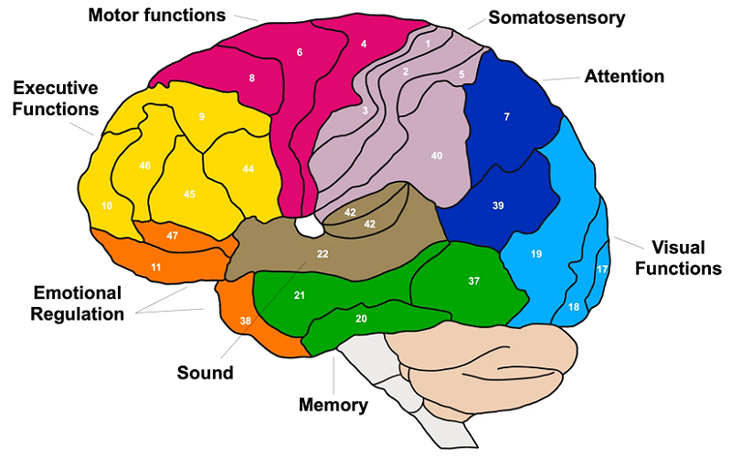
Area 1, 2, and 3: Primary Somatosensory Cortex
Nestled in the postcentral gyrus of the parietal lobe, these three regions collectively form the primary somatosensory cortex. This is where the initial cortical processing of tactile sensations, like touch, temperature, and proprioception (awareness of body position), occurs.
- Area 1 is predominantly concerned with processing tactile texture and shape.
- Area 2 deals with size and shape recognition, integrating touch with proprioceptive information.
- Area 3 can be further subdivided into Area 3a and 3b. While 3a processes proprioceptive information, 3b is responsible for basic tactile sensations.
Together, these areas paint a vivid picture of our physical interactions with the world, ensuring we can sense, respond to, and navigate our environment.
Area 4: Primary Motor Cortex
Positioned anteriorly to the somatosensory areas in the precentral gyrus, Area 4 plays a pivotal role in voluntary motor control. It generates neural impulses that control the execution of movement. Neurons in this area project directly to the spinal cord and brainstem, influencing motor neurons and ultimately determining muscle contractions. Interestingly, certain parts of the body, like the hands and face, have a larger representation in the primary motor cortex due to their intricate motor needs [3].
Area 17: Primary Visual Cortex
Tucked away in the depths of the occipital lobe, Area 17, often called V1, stands as the gateway to visual perception. All visual information relayed from our eyes first lands here. V1 processes basic visual features like edge detection, light contrast, and orientation. It’s from this foundational processing that all other visual experiences, including recognition of objects, faces, or places, are constructed in associated visual areas.
Area 41 and 42: Primary Auditory Cortex
Situated within the temporal lobe, these areas serve as the central hub for auditory processing. Sound waves captured by our ears are transformed into electrical signals, which are then processed and interpreted in these regions.
- Area 41 is primarily responsible for the basic auditory input, decoding parameters like pitch and volume.
- Area 42 takes on a more integrative role, assisting in the recognition of complex sounds such as melodies or familiar voices.
Other Notable Areas and Their Significance
While the aforementioned regions have clear sensory or motor roles, many Brodmann Areas contribute to higher cognitive functions, emotions, and more complex integrations:
- Areas 9, 10, 11 are located in the prefrontal cortex, these areas play roles in planning, decision-making, and personality expression.
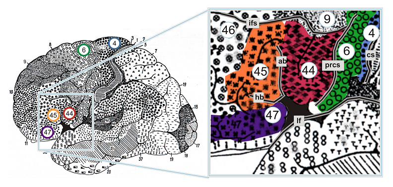
- Areas 44 and 45 are often referred to as Broca’s region, these areas in the frontal lobe are crucial for speech production and some aspects of language comprehension.

Importance of Brodmann Areas in Modern Neuroscience
Even as we’ve journeyed deep into the 21st century, with its wealth of technological advancements and newfound scientific paradigms, the work of Korbinian Brodmann continues to echo throughout the hallways of neuroscience. The conceptualization of Brodmann Areas has endured not merely as a historical curiosity but as a foundational framework, reinforcing and guiding modern neurological research.
Role in Functional MRI (fMRI) Studies
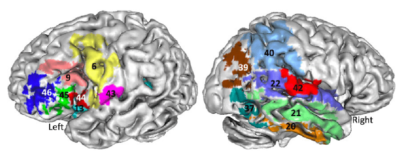
Functional Magnetic Resonance Imaging, or fMRI, has revolutionized our ability to observe the active brain in real-time. By measuring changes in blood flow, fMRI provides insights into which parts of the brain are engaged during specific tasks or cognitive processes [4].
Brodmann Areas serve as a ready reference in these studies. When researchers observe activation in a specific cortical region, it’s often described in terms of the corresponding Brodmann Area. This provides a universally understood language, ensuring clear communication across the global scientific community. Additionally, the distinct cytoarchitectural properties of these areas can influence blood flow patterns, which in turn affects fMRI signal and interpretation.
Understanding Neurological Disorders
Numerous neurological and psychiatric disorders manifest through structural and functional anomalies in specific cortical regions. Brodmann Areas offer a precise way to pinpoint these anomalies. For instance:
- In Alzheimer’s disease, degeneration often begins in Area 24, impacting the anterior cingulate cortex.
- Schizophrenia has been linked to abnormalities in several Brodmann Areas, including Areas 9 and 10, which are associated with higher cognitive functions.
By understanding the typical functions of these affected areas, researchers and clinicians can better predict, diagnose, and potentially treat the associated symptoms.
Impact on Cognitive Science and Psychology
Beyond the boundaries of pure neurology, Brodmann’s legacy has deeply influenced the fields of cognitive science and psychology. Cognitive tasks often involve a network of brain regions working in concert. By understanding the Brodmann Areas involved, researchers can draw clearer connections between structure and function, linking neural activity to specific cognitive or psychological phenomena.
For instance, studies on memory might reference activations in Areas 28 and 34, parts of the entorhinal cortex crucial for memory encoding and recall. Meanwhile, investigations into emotion regulation might highlight the role of Area 25, the subgenual cingulate, known to play a part in mood disorders.

Brodmann Areas Controversies and Limitations
Like any pioneering scientific work, Brodmann’s classification of cortical areas has faced its fair share of scrutiny, debate, and evolution. While the importance of these areas in neuroscientific research is undeniable, it’s also essential to approach them with an awareness of the limitations and controversies they carry. This nuanced perspective ensures that we remain open to new findings and interpretations, always pushing the boundaries of knowledge.
Individual Variability
One of the most significant challenges in generalizing Brodmann’s findings is the recognition that there is substantial individual variability in brain anatomy and function. While his areas provide a generalized map.
Anatomical Variations
The exact borders and sizes of Brodmann Areas can vary from person to person. For instance, the renowned Area 44, often associated with speech production, can manifest differently across individuals, making it challenging to pinpoint in some cases.
Functional Variations
Even if the anatomical boundaries are consistent, the functions associated with a given Brodmann Area might differ among people, especially when considering left-handed versus right-handed individuals or those with atypical neural wirings [5].
Over-reliance on Cytoarchitecture
While Brodmann’s classification was primarily based on cytoarchitecture, modern neuroscience has shown that structure and function don’t always align neatly:
Functional Overlaps
A single Brodmann Area can play roles in multiple functions. For example, Area 6 is involved in both motor planning and cognitive processes, blurring the boundaries of strict functional categorization.
Structural Subdivisions
Advances in microscopy and imaging have revealed that some of Brodmann’s areas can be further subdivided based on even finer structural details or functional differentiations.
The Debate on Exact Numbers
While Brodmann initially identified 52 distinct areas, subsequent research, equipped with more advanced tools and methodologies, has debated this number. Some researchers argue that there are fewer areas, suggesting that some of Brodmann’s regions can be combined based on similar characteristics. In contrast, others believe there are even more nuanced divisions, extending beyond the original 52.
The Evolutionary Perspective
Human brains aren’t static entities. They’ve evolved over millions of years, adapting to changing environments and needs. Some critics argue that Brodmann’s areas, while accurate for the time, might not fully encompass the evolutionary adaptations and complexities present in modern humans.
References
[1] Brodmann Areas: Anatomy And Functions
[2] Brodmann: a pioneer of human brain mapping—his impact on concepts of cortical organization
[3] Centenary of Brodmann’s map — conception and fate
[4] Architectonic Mapping of the Human Brain beyond Brodmann
[5] Brodmann’s cortical maps

