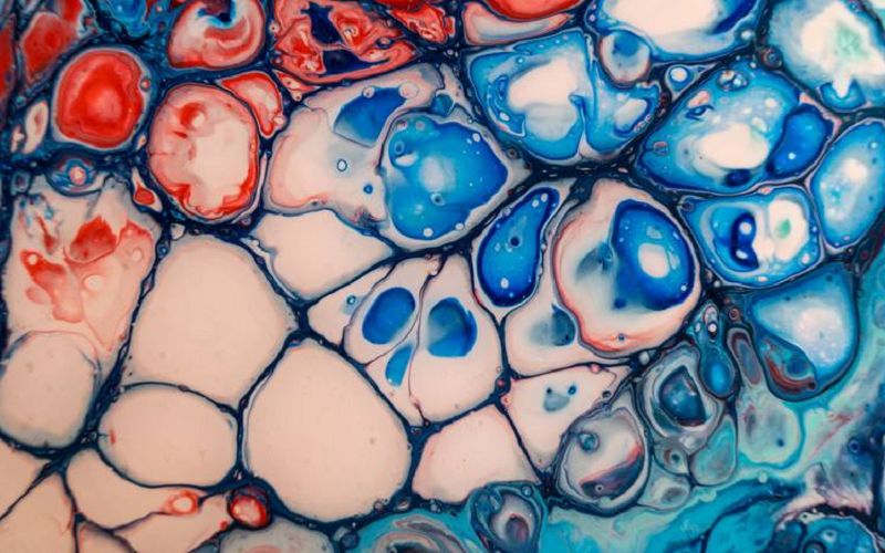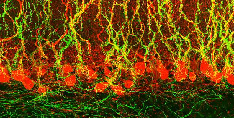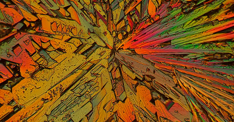
In the vast realm of art, where expansive canvases and monumental sculptures often claim the limelight, there lies a unique, intricate world that commands our attention in a subtly powerful way: microscopic art. This art form, characterized by its minute details and mesmerizing patterns, isn’t just a visual marvel; it holds a mirror to the intricate designs of our very own neural pathways. This interplay between what we observe externally and its influence on the internal wiring of our brains offers a captivating narrative.
Contents
- Introduction to Microscopic Art and Neural Patterns
- History of Microscopic Art
- The Purkinje Pattern and Neural Patterns
- How Microscopic Art Stimulates the Brain
- Neural Plasticity and Adaptation
- References
Introduction to Microscopic Art and Neural Patterns
In the vast canvas of artistic expression, there exists a domain often overlooked by the casual observer yet profoundly impactful in its allure: microscopic art. This diminutive yet dazzling form of artistic endeavor offers more than just visual splendor—it intertwines with the very fabric of our cognitive processes, influencing the patterns and rhythms of our neural networks.
Definition of Microscopic Art
Microscopic art, as the name suggests, is an art form that is typically observed and appreciated at minuscule scales. This does not necessarily mean the art is created using microscopes, though it often is; rather, it signifies art that showcases incredibly detailed and precise work, often mimicking or reflecting patterns seen at the microscopic level in nature. From depictions of cellular structures to the minute patterns of crystalline formations, this art form captivates by presenting the unseen wonders of our universe.
Brief Overview of Neural Patterns
To fully grasp the connection between microscopic art and our brain, it’s essential to understand neural patterns. Neural patterns, or neural circuits, are the pathways formed by interconnected neurons in the brain. These pathways are responsible for transmitting and processing information. Think of them as the highways of the brain, guiding our thoughts, emotions, and actions. Over time, with repetition and experience, certain neural patterns become strengthened, influencing our habits, behaviors, and even our perception of the world.
Purpose and Importance of Studying the Relationship
The interplay between microscopic art and neural patterns is not a mere coincidence. As humans, we are naturally drawn to patterns, symmetry, and detail—attributes that are abundant in microscopic art. By exploring this relationship, we can gain insights into how visual stimuli, especially detailed and intricate forms like microscopic art, can influence our brain’s functioning, potentially offering therapeutic benefits and enhancing cognitive health. This union of art and neuroscience provides a novel lens through which we can view and appreciate the profound impacts of art on the human psyche [1].
History of Microscopic Art
While the concept of art and artistic expression is as old as humanity itself, the foray into the intricate realm of microscopic art is a fascinating journey spanning centuries, evolution, and technological advancements. This journey not only showcases the sheer skill of artists but also the human propensity to find beauty in the minutiae and to depict the wonders that the naked eye might miss.
Evolution of Microscopic Art Over the Years
Microscopic art, in its nascent stages, wasn’t necessarily about depicting the microscopic, but rather about the painstaking precision required to produce incredibly detailed works on minuscule canvases. Think of ancient inscriptions on coins, minute paintings on grains of rice, or the intricate designs of some indigenous jewelry.
As science and art began to intertwine with the invention and popularization of the microscope in the 17th century, artists found inspiration in the previously unseen microscopic world. This newly accessible realm of bacteria, blood cells, and crystalline structures opened a door to an entirely new genre of artistic representation.
Famous Microscopic Artists and Their Contributions
Over the years, several artists have distinguished themselves in the field of microscopic art, each contributing a unique perspective.
Robert Hooke
Often credited as one of the pioneers in this domain, Hooke’s seminal work, “Micrographia,” published in 1665, offered detailed sketches of the microscopic realm, from insects to plant cells. It was not just a scientific record but also a testament to the intricate beauty of the micro-world.
Klaus Kemp
Known as the modern master of diatom arrangement, Kemp meticulously arranges microscopic algae called diatoms into kaleidoscopic patterns, reviving a Victorian art form and showcasing the beauty of these minute organisms.
Willard Wigan
An artist renowned for his micro-sculptures, Wigan creates art so tiny it often needs to be viewed under a microscope. From sculptures inside the eye of a needle to art on a human hair, his works challenge our perceptions of scale and detail.
Technological Advances Aiding in the Creation of Microscopic Art
The progression of microscopic art would be incomplete without acknowledging the role of technology. As microscopes evolved, offering clearer, magnified images with higher resolutions, artists were able to delve deeper into the world of the minute. Today, advancements in digital imaging and electron microscopy provide artists not only with inspiration but also with tools.
These technological marvels allow for manipulation at a microscopic scale, letting artists create in spaces and with materials that were once deemed impossible. From using 3D printing at a micro-level to manipulating cells to create bio-art, the boundaries of microscopic art continue to expand with technological innovation [2].

The Purkinje Pattern and Neural Patterns
The Purkinje Pattern, also referred to as the Purkinje Shift, is a physiological phenomenon observed in the human visual perception system. It pertains to the shift in peak sensitivity of the human eye from the green wavelengths to the blue wavelengths as the lighting transitions from daylight to twilight. Named after the Czech physiologist Jan Evangelista Purkinje, who discovered it in the 19th century, this shift is attributed to the differing functionalities of the cone and rod cells in the human retina.
While cone cells are primarily responsible for color vision under well-lit conditions, rod cells dominate under low light conditions and are most sensitive to blue-green wavelengths. As the ambient light decreases, the eye’s sensitivity shifts towards the spectral range where rods are more active, thus the perceived color emphasis transitions from greens to blues. This phenomenon underpins the challenges in achieving consistent color perception across varying light conditions and has implications in fields like photography, lighting design, and astronomy.
How the Purkinje Pattern Related to Neural Patterns
The Purkinje Pattern, in its essence, is a manifestation of the underlying neural patterns and mechanisms of the human visual system. Neural patterns refer to the organized and consistent ways in which neurons in the brain and nervous system respond to specific stimuli or perform certain functions [3]. Here’s how the Purkinje Pattern relates to these neural patterns.
Differential Activation of Photoreceptors
The human retina contains two primary types of photoreceptors: rods and cones. Each has a distinct neural response to different wavelengths of light. The Purkinje Shift arises from the differential sensitivity and neural activation patterns of these photoreceptors under varying light conditions.
Adaptation Mechanisms
Neural adaptation is a fundamental property of neural systems where the responsiveness of neurons changes as a result of previous stimuli. In the context of the Purkinje Shift, as lighting conditions change, the neural responses of the visual system adapt, leading to a shift in color sensitivity.
Processing in the Retina and Brain
Once light is detected by photoreceptors, the information is processed by retinal ganglion cells and other intermediate neurons before being sent to the brain. The way these neurons process and relay information can influence color perception, and these processing patterns play a role in phenomena like the Purkinje Shift.
Interaction with Cognitive Processes
While the Purkinje Pattern primarily stems from physiological processes, the brain’s interpretation of visual information is also influenced by cognitive processes, memories, and expectations. Neural patterns in higher-order visual processing centers in the brain can further modulate the perception of colors under different lighting.

How Microscopic Art Stimulates the Brain
Art has always been a conduit for human emotion, thought, and perception. Yet, microscopic art, with its intricate designs and almost otherworldly depictions, offers a unique cerebral experience.
Visual Processing of Intricate Details
As one gazes upon a piece of microscopic art, the initial reaction is often one of wonder. This reaction is not just emotional but is deeply rooted in the intricate processes of our visual system.
Engagement of the Occipital Lobe
The primary visual cortex, located in the occipital lobe, is the first to process the visual information. Here, the intricate details, lines, and contrasts found in microscopic art are broken down, analyzed, and interpreted.
Perception of Colors, Shapes, and Patterns
As the brain continues to process the artwork, other regions get involved. The fusiform gyrus, for instance, assists in recognizing specific shapes and patterns, while the color centers of the brain revel in the vibrant hues and subtle gradients that microscopic art often presents. These processes allow us to appreciate the artistry and precision involved [4].
Cognitive Processing and Interpretation
Beyond just visual processing, microscopic art demands a level of cognitive engagement that is both intriguing and enlightening.
Activation of the Prefrontal Cortex
As we try to understand and interpret the often abstract nature of microscopic art, our prefrontal cortex – responsible for complex cognitive behavior, decision-making, and moderating social behavior – springs into action. We begin to contextualize the artwork, drawing from our experiences, memories, and knowledge.
Involvement of Memory and Past Experiences
The hippocampus, central to memory formation and retrieval, plays a role when familiar patterns or subjects are depicted in the art. For instance, a piece showcasing a magnified leaf structure might evoke personal memories of nature walks or forest adventures, adding layers of personal connection to the viewing experience.
Neural Plasticity and Adaptation
One of the marvels of the human brain is its ability to adapt and reorganize itself, especially in response to new experiences. Engaging with microscopic art is no exception.
Repetitive Engagement and Strengthening Neural Pathways
The more we expose ourselves to the detailed wonders of microscopic art, the stronger the associated neural pathways become. Much like learning a musical instrument or a new language, repetitive engagement enhances synaptic strength, allowing for quicker and richer processing of similar stimuli in the future.
Novelty and the Brain’s Reward System
Our brains are hardwired to enjoy and seek out novel experiences. Microscopic art, given its unique nature, often presents imagery and details that are novel to the viewer. This activates our brain’s reward system, releasing neurotransmitters like dopamine, which induce feelings of pleasure and satisfaction.
Neural Plasticity and Adaptation
At the heart of understanding our brain’s interaction with microscopic art is the fascinating phenomenon of neural plasticity. This dynamic ability of the brain to reorganize and adapt itself is both its superpower and its subtle response mechanism. Microscopic art, with its depth and intricacy, has the potential to play a significant role in influencing and being influenced by this neural adaptability.
Introduction to Neural Plasticity
Neural plasticity, often referred to as neuroplasticity, is the brain’s remarkable capability to reconfigure its connections and pathways based on experiences, learning, and environmental influences. Contrary to once-popular belief that the brain’s structure is static post-childhood, research over the past few decades has illuminated the brain’s continuous ability to adapt, even into adulthood. This adaptability can be witnessed in various forms, from the formation of new synapses to the strengthening or weakening of existing neural connections.
How Repetitive Viewing Can Shape Neural Pathways
Repeated experiences, including consistent exposure to certain stimuli like art, can have a lasting impact on our brain’s structure and function. Microscopic art, given its detailed nature, offers a particularly engaging stimulus [5].
Strengthening of Synaptic Connections
Just as a musician might strengthen neural pathways related to hand-eye coordination or auditory processing through practice, frequent engagement with microscopic art can solidify the neural pathways associated with visual processing and cognitive interpretation of such intricate details.
Formation of New Neural Pathways
Beyond just strengthening existing connections, new experiences with varied pieces of microscopic art can lead to the formation of entirely new pathways. This essentially means that as we expose ourselves to more diverse microscopic artworks, our brain continually adapts, creating fresh networks to process and appreciate these novel stimuli.
The Role of Curiosity and Exploration in Brain Health
Intrinsic to the experience of delving into microscopic art is a sense of curiosity and exploration. This feeling isn’t just a fleeting emotion; it has tangible neural implications.
Activation of the Dopaminergic System
Curiosity, as a driving force, activates the brain’s dopaminergic system, a network deeply involved in reward, pleasure, and motivation. As we satiate our curiosity by exploring microscopic art, dopamine is released, reinforcing our desire to learn and explore further.
Enhanced Learning and Memory
A brain fueled by curiosity is more receptive to learning. When engaging with microscopic art, this heightened state of curiosity can make us more attuned to details, aiding in better retention and deeper appreciation of the artwork.
Resilience and Adaptability
Consistently challenging our brains through exploration and novel experiences, like deciphering intricate art pieces, contributes to long-term brain health. It fosters resilience against cognitive decline and encourages a mindset of adaptability and continuous learning.
References
[1] Spectacular Microscopic Art Is Also World-Changing Science
[2] Dazzling Images of the Brain Created by Neuroscientist-Artist
[3] The Purkinje Pattern
[4] Art That Gets You Thinking
[5] Etching the Neural Landscape

